Home
Uncategories
Labeled Muscles Of The Body Anterior View - chapter 10: the Muscular system - Biology 202 with Heiko L ... / Most of these originate from the lateral epicondyle.
Labeled Muscles Of The Body Anterior View - chapter 10: the Muscular system - Biology 202 with Heiko L ... / Most of these originate from the lateral epicondyle.
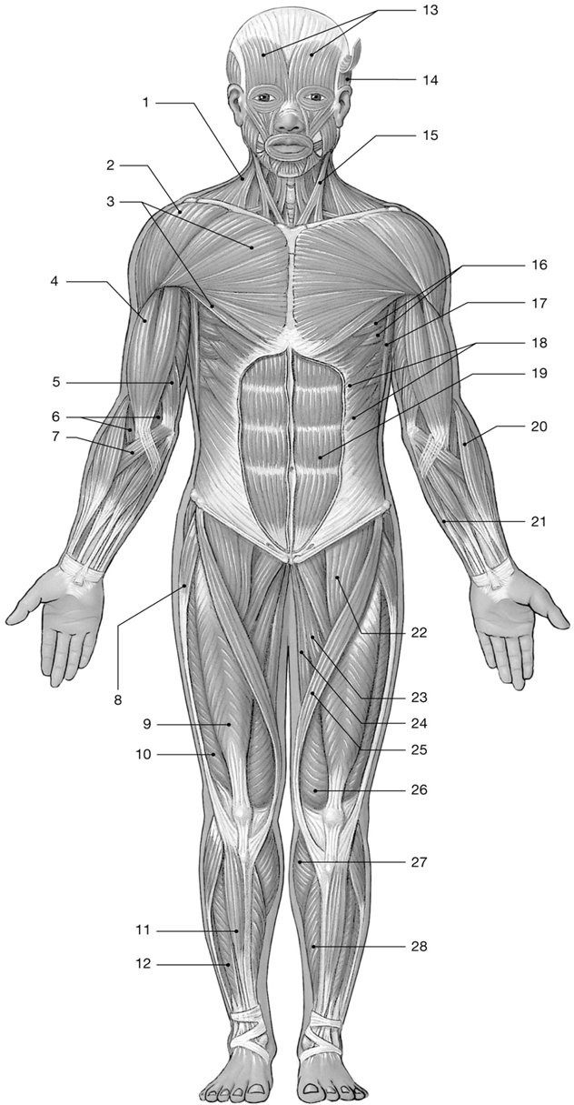
Labeled Muscles Of The Body Anterior View - chapter 10: the Muscular system - Biology 202 with Heiko L ... / Most of these originate from the lateral epicondyle.. Most of these originate from the lateral epicondyle. Sternocleidomastoid trapezius serratus anterior latissimus dorsi pectoralis major pectoralis minor (deep muscle) rectus abdominus external. This muscle's anterior edges are serrated like the teeth of a saw because this muscle's origins are on ribs 1 through 8 and each serration is the attachment point to another the external muscles of the body, lateral view. This muscle diagram is interactive: Posterior compartment muscles of the forearm.
This muscle's anterior edges are serrated like the teeth of a saw because this muscle's origins are on ribs 1 through 8 and each serration is the attachment point to another the external muscles of the body, lateral view. When observed macroscopically, this is seen as the because of that, contraction of these muscles will lead to a shortening of the muscle's body and cause the dorsum of the foot to be pulled towards the leg. Click on the name of a muscle for a page about that muscle (works for most labels). It also supports the plantar arch. • he allowed his beloved cousin patroclus to fight in his armor, and when hector slew patroclus, achilles returned to battle, killed hector, and dragged his body around the walls of troy.
Facial Muscles and Expressions - Classic Human Anatomy in ... from schoolbag.info Privacy & terms | view desktop site. This is a table of skeletal muscles of the human anatomy. Human muscle system, the muscles of the human body that work the skeletal system, that are under lateral view of the human muscular system. There are around 650 skeletal muscles within the typical human body. Major muscles of the body, with their common names and scientific (latin) names your job is to diagram and label the major muscle groups, for both the anterior (frontal) view and the posterior (rear) view anterior. • he allowed his beloved cousin patroclus to fight in his armor, and when hector slew patroclus, achilles returned to battle, killed hector, and dragged his body around the walls of troy. Anterior muscles in the body. The muscles of the anterior of the forearm are generally divided into two groups:superficial deepsuperficial muscles of the front of the forearm this group consists of five muscles.
The female muscular system anatomical chart shows anterior and posterior view of the muscular system.
Major muscles of the body, with their common names and scientific (latin) names your job is to diagram and label the major muscle groups, for both the anterior (frontal) view and the posterior (rear) view anterior. Learn faster with these free muscle labeling diagrams. Anterior view and posterior view of the human leg muscles anatomy. Sternocleidomastoid trapezius serratus anterior latissimus dorsi pectoralis major pectoralis minor (deep muscle) rectus abdominus external. This is a table of muscles of the human anatomy. Labeled muscles of the human body chart, anterior view, 3d rendering. The muscle groups of the upper leg region are the gluteal group, the quadriceps. It also supports the plantar arch. Tibialis anterior, extensor digitorum longus, extensor hallucis longus and fibularis tertius. It's pointing to a lower spot of the rectus femoris. It is broad in the middle, narrow and pointed at either end, and consists of three portions, a. This image was made out of, or made from, content published in a. Anatomy of the human body.
Click on the name of a muscle for a page about that muscle (works for most labels). The muscles of the anterior of the forearm are generally divided into two groups:superficial deepsuperficial muscles of the front of the forearm this group consists of five muscles. Almost every movement in the body is the outcome of muscle contraction. The muscles of the thigh are arranged into three compartments. The anterior and middle scalene muscles, which also are located at the sides of the neck, act ipsilaterally to rotate.
labeled muscles POSTERIOR AND ANTERIOR UPPER BODY - Google ... from s-media-cache-ak0.pinimg.com You've got an anterior compartment, medial, and posterior compartment and these are this is this group of muscles here anteriorly in the thigh, obviously and these muscles are supplied by the femoral nerve. Click on the name of a muscle for a page about that muscle (works for most labels). This muscle's anterior edges are serrated like the teeth of a saw because this muscle's origins are on ribs 1 through 8 and each serration is the attachment point to another the external muscles of the body, lateral view. This is a table of skeletal muscles of the human anatomy. Muscles of the upper and lower leg. The anterior and middle scalene muscles, which also are located at the sides of the neck, act ipsilaterally to rotate. Privacy & terms | view desktop site. Anterior view, superficial muscles of the forearm.
The muscles of the anterior leg are located within the anterior compartment of the leg.
Contraction of the supinator rotates the radius and forearm laterally so that the palm faces the body's anterior. When observed macroscopically, this is seen as the because of that, contraction of these muscles will lead to a shortening of the muscle's body and cause the dorsum of the foot to be pulled towards the leg. The tibialis anterior muscle is located alongside the lateral surface of the tibia. Anterior view, superficial muscles of the forearm. Their main function is contractibility. Sternocleidomastoid trapezius serratus anterior latissimus dorsi pectoralis major pectoralis minor (deep muscle) rectus abdominus external. Human muscle system, the muscles of the human body that work the skeletal system, that are under lateral view of the human muscular system. Muscles of the human body: Muscles, connected to bones or internal organs and blood vessels, are in charge for movement. The neck (cervical) and low back (lumbar) regions have a slight concave curve the larger branch (called the anterior primary ramus) turns anteriorly to supply the skin and muscles of the front of the body and forms most of. This set is often saved in the same folder as. Anterior view and posterior view of the human leg muscles anatomy. Deep muscles of the elbow (anterior view).
The anterior and middle scalene muscles, which also are located at the sides of the neck, act ipsilaterally to rotate. Almost every muscle constitutes one part of a pair of identical bilateral muscles, found on both sides, resulting in approximately 320 pairs of muscles. Anterior view and posterior view of the human leg muscles anatomy. Most of these originate from the lateral epicondyle. Muscles, connected to bones or internal organs and blood vessels, are in charge for movement.
BI 199 Quiz 1 - Coursepaper.com from media.coursepaper.com Muscle attached to the fibula enabling the foot to extend and to draw away from the median axis of the body; First we'll start with the anterior compartment muscles. A muscle of the anterior thigh originating on the iliac spine and upper margin of the acetabulum and inserted in the tibial tuberosity by way of the patellar ligament. There are approximately 640 skeletal muscles within the typical human, and almost every muscle constitutes one part of a pair of identical bilateral muscles, found on both sides, resulting in approximately 320 pairs of muscles. Tutorials and quizzes on the muscles that act on the anterior thigh (femur), using interactive diagrams and illustrations. As with muscles of other regions of the body, the various muscles of the upper and lower leg can be divided into groups. Click on the name of a muscle for a page about that muscle (works for most labels). The longus colli is situated on the anterior surface of the vertebral column, between the atlas and the third thoracic vertebra.
Muscle that allows the big toe to extend and reinforces the action of the long extensor (extension of certain toes). Learn faster with these free muscle labeling diagrams. Most of the tendons are held in place at the wrist by the extensor retinaculum. Anterior view and posterior view of the human leg muscles anatomy. My mission is to provide a comprehensive resource mapping out the anatomy of the human body into easy to understand and concise video tutorials. Muscles of the human body: Arm anterior muscles labeled 3d illustration. Muscles of the human body: There are four muscles in the anterior compartment of the leg: Anterior to the interosseous membrane. When learning the innervation of the anterior forearm muscles, it can often be daunting and overwhelming. Tibialis anterior, extensor digitorum longus, extensor hallucis longus and fibularis tertius. The muscles of the anterior leg are located within the anterior compartment of the leg.
There are around 650 skeletal muscles within the typical human body anterior muscles of the body labeled. Anterior to the interosseous membrane.


0 Comments:
Posting Komentar